Vascular calcification doesn’t announce itself. It does not cause pain or a tingling feeling. The arteries become stiffer and narrower without any signs until an MRI or heart condition is detected. For those suffering from diabetes, the disease progresses much faster due to constant high levels of glucose and abnormal mineral metabolism. Diabetology programs always pay close attention to glucose, blood pressure, and cholesterol levels. Much less often, there is monitoring of the protein responsible for blocking calcium from arteries and vitamin supporting its activity.
The Protein Standing Between Calcium and Your Arteries
Matrix Gla protein (MGP) is the body’s primary defense against calcium accumulating where it shouldn’t. It only works once carboxylated which is a step for which vitamin K is the required cofactor. Mice bred without functional MGP develop normally to birth but die within two months from catastrophic arterial calcification and vessel rupture (Luo et al., 1997). In humans the process is slower, but the mechanism is the same that without enough active vitamin K, MGP circulates in an inactive form that can’t do its job.
What a Cohort of 518 Diabetics Showed
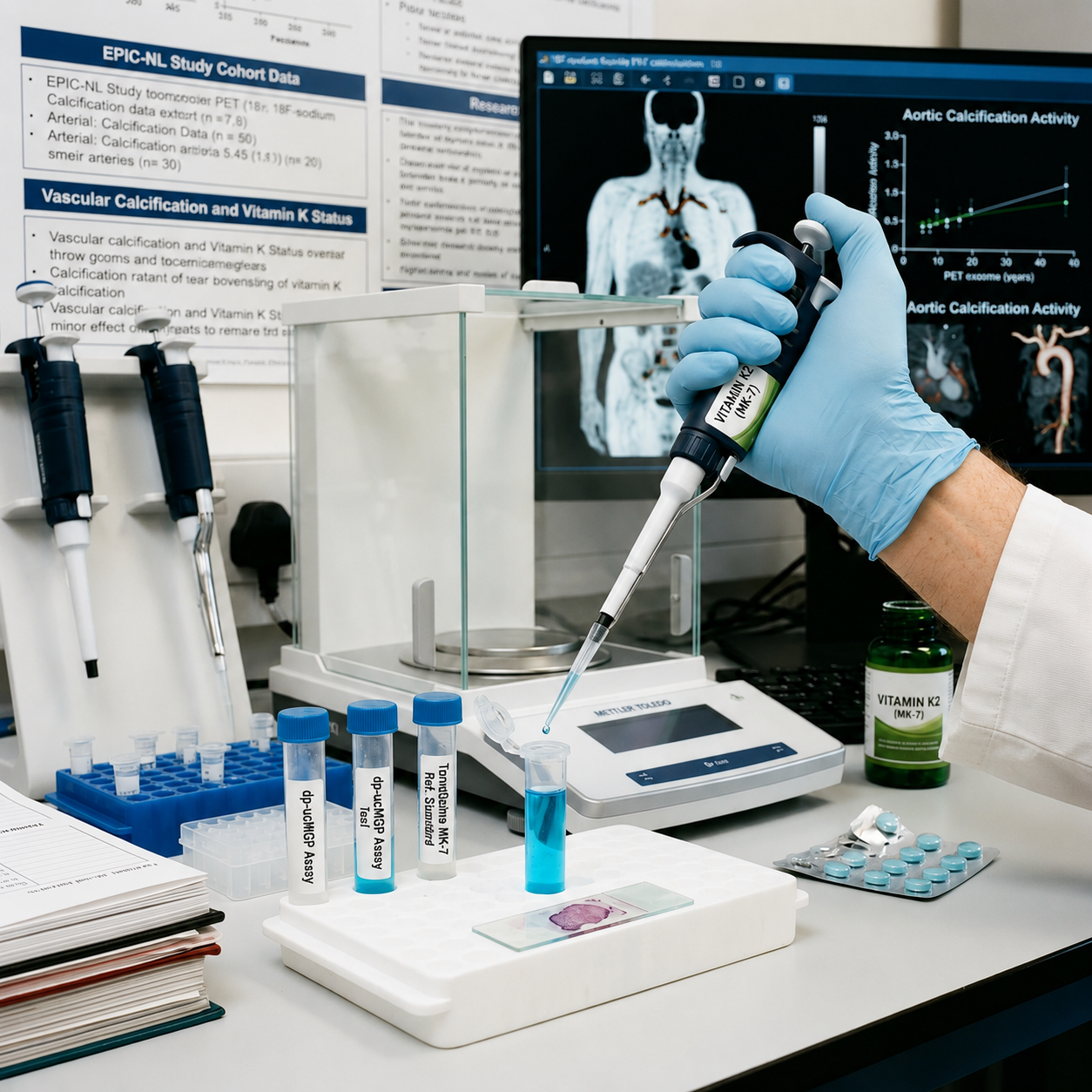
A Dutch cohort study followed 518 people with type 2 diabetes as part of the larger EPIC-NL study, measuring circulating MGP species at baseline and tracking cardiovascular events over the following years (Dalmeijer et al., 2013). Patients with high levels of dp-ucMGP which is the inactive, uncarboxylated form that signals poor vitamin K status had significantly higher rates of cardiovascular disease, particularly peripheral arterial disease and heart failure. Other circulating MGP forms weren’t linked to risk the same way; it was specifically the inactive fraction that mattered. The researchers’ conclusion was direct that poor vitamin K status tracked with cardiovascular risk in exactly the population where vascular calcification tends to progress fastest.
Calcification in Real Time
Most calcification research measures static calcium scores which is like a snapshot of damage already done. A 2022 trial did something different: it used 18F-sodium fluoride PET imaging, which lights up sites of active, ongoing calcification rather than calcium that has already settled in place (Bellinge et al., 2022). Diabetic patients with existing coronary calcium scores above 10 took either 10 milligrams of vitamin K1 daily or a placebo for three months, then were re-scanned to see where new calcification activity had appeared:
| Site (18F-NaF PET scan) | Odds of new active calcification vs. placebo | Significance |
| Coronary arteries | 65% lower odds | OR = 0.35, p = .010 |
| Aorta | 73% lower odds | OR = 0.27, p = .040 |
| Coronary + aortic combined | 72% lower odds | OR = 0.28, p = .002 |
Source: Bellinge, Francis, Lee, Bondonno, Sim, Lewis, Watts, & Schultz (2022).
The Bigger Evidence Base
A single PET-imaging trial deserves context. A 2023 meta-analysis pooling 14 randomized trials and 1,533 patients found that vitamin K supplementation significantly slowed progression of coronary artery calcium scores overall (mean difference −17.37, p = .04) (Li et al., 2023). The population isn’t diabetes-specific, but it points the same direction that adequate vitamin K status appears to measurably slow arterial calcification, not just theoretically inhibit it.
Why the Form of K2 Matters for a Slow-Moving Process
Calcium deposition takes place over years rather than days, therefore the constant state of vitamin K becomes important in its efficacy rather than the dose taken alone. One study on the bioavailability of MK-7 showed that it was still detected in the bloodstream up to 48 hours post one dosage, while MK-4, taken in an equivalent amount, was undetectable at any time period (Sato et al., 2012). It seems reasonable to conclude that a version of K2 that maintains consistent blood concentration daily would be more suitable.
Where TerraQuino Fits In
TerraQuino produces MK-7 through a controlled fermentation process, delivering the long-chain, allergen-free vitamin K2-7 associated with the sustained blood levels documented above. It’s formulated for tablets, capsules, softgels, sachets, and liquids, and pairs cleanly with vitamin D3, calcium, and omega-3s, common in heart-health formulations.
TerraQuino doesn’t make disease-specific claims for its ingredient; the diabetes-focused trials above used vitamin K1 or measured general MGP status rather than testing MK-7 directly. It gives formulators a bioavailability-tested K2 source to build toward that evidence base.
Frequently Asked Questions
Does vitamin K2 reverse arterial calcification that’s already there?
Not established. The clearest trial evidence shows vitamin K reducing new, active calcification (Bellinge et al., 2022) and slowing calcium-score progression (Li et al., 2023), not reversing plaque that’s already calcified.
Why hasn’t my doctor mentioned this?
Vitamin K’s role in vascular calcification is a newer, smaller research area than blood sugar or cholesterol management, and most trials so far are modest in size. It hasn’t yet made it into standard diabetes care guidelines.
Is vitamin K1 or K2 better studied for this specific effect?
The direct diabetic-population RCT used K1 (Bellinge et al., 2022). MGP-status research in diabetics has focused on overall vitamin K carboxylation activity rather than K1 versus K2 specifically (Dalmeijer et al., 2013). MK-7 is generally favored in supplements for its longer half-life (Sato et al., 2012).
Can I take vitamin K2 if I’m on blood thinners?
Vitamin K interacts directly with warfarin and similar anticoagulants. Anyone on blood-thinning medication should talk to their prescribing doctor before adding any form of vitamin K.
References
Bellinge, J. W., Francis, R. J., Lee, S. C., Bondonno, N. P., Sim, M., Lewis, J. R., Watts, G. F., & Schultz, C. J. (2022). The effect of vitamin K1 on arterial calcification activity in subjects with diabetes mellitus: A post hoc analysis of a double-blind, randomized, placebo-controlled trial. The American Journal of Clinical Nutrition, 115(1), 45–52. https://doi.org/10.1093/ajcn/nqab306
Dalmeijer, G. W., van der Schouw, Y. T., Magdeleyns, E. J., Vermeer, C., Verschuren, W. M., Boer, J. M., & Beulens, J. W. (2013). Matrix Gla protein species and risk of cardiovascular events in type 2 diabetic patients. Diabetes Care, 36(11), 3766–3771. https://doi.org/10.2337/dc13-0065
Li, T., Wang, Y., & Tu, W. (2023). Vitamin K supplementation and vascular calcification: A systematic review and meta-analysis of randomized controlled trials. Frontiers in Nutrition, 10, Article 1115069. https://doi.org/10.3389/fnut.2023.1115069
Luo, G., Ducy, P., McKee, M. D., Pinero, G. J., Loyer, E., Behringer, R. R., & Karsenty, G. (1997). Spontaneous calcification of arteries and cartilage in mice lacking matrix GLA protein. Nature, 386(6620), 78–81. https://doi.org/10.1038/386078a0
Sato, T., Schurgers, L. J., & Uenishi, K. (2012). Comparison of menaquinone-4 and menaquinone-7 bioavailability in healthy women. Nutrition Journal, 11, Article 93. https://doi.org/10.1186/1475-2891-11-93